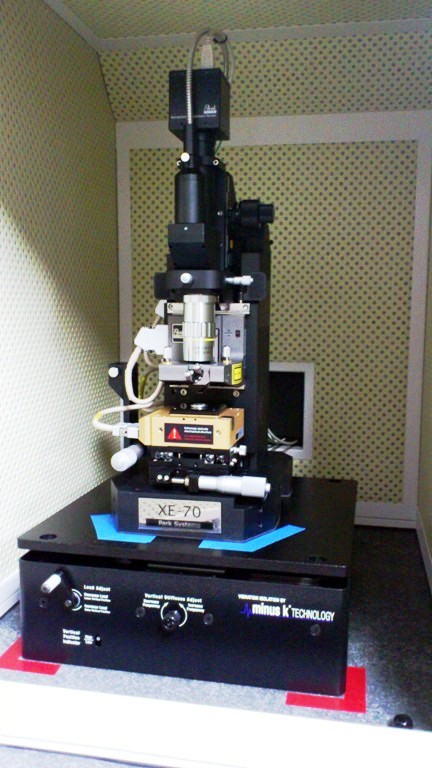
Click for more images: Atomic Force Microscope (AFM)

Atomic force microscopy (AFM) or scanning force microscopy (SFM) is a very high-resolution type of scanning probe microscopy, with demonstrated resolution on the order of fractions of a nanometer, more than 1000 times better than the optical diffraction limit. The precursor to the AFM, the scanning tunneling microscope, was developed by Gerd Binnig and Heinrich Rohrer in the early 1980s at IBM Research – Zurich, a development that earned them the Nobel Prize for Physics in 1986. Binnig invented the atomic force microscope (also abbreviated as AFM) and the first experimental implementation was made by Binnig, Quate and Gerber in 1986. The first commercially available atomic force microscope was introduced in 1989. The AFM is one of the foremost tools for imaging, measuring, and manipulating matter at the nanoscale. The information is gathered by “feeling” the surface with a mechanical probe. Piezoelectric elements that facilitate tiny but accurate and precise movements on (electronic) command enable the very precise scanning. In some variations, electric potentials can also be scanned using conducting cantilevers. In more advanced versions, currents can be passed through the tip to probe the electrical conductivity or transport of the underlying surface, but this is much more challenging with few research groups reporting consistent data (as of 2004).
(Source: Wikipedia)
Click for more images: Atomic Force Microscope (AFM)
How AFM Works: AFM Principle
http://www.parkafm.com/index.php/medias/nano-academy/how-afm-works
AFM Modes and Techniques
http://www.parkafm.co.kr/index.php/kr/products/modes-options/park-afm-modes
(Source: Park Systems)
